Global Interventional Radiology
Precision Imaging & Clinical Interventions
Access the latest research, procedural guidelines, and complex case studies curated for the global interventional radiology community.
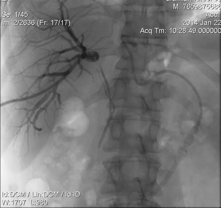
01. Portography after left PV peripheral patentbranch puncture 02. Introducer sheath is advance and portography “above the thrombus” is performed 03. Portography “below the thrombus” – tumor thrombus is documented 04. Guidewire and 5Fr diameter advantage catheter is introduced in left gastric vein (LGV) 05. Contrast injection in LGV depicts esophageal varicosis 06. LGV embolization by coil implantation 07. Post-embolization contrast injection in LGV documents no more esophageal varicosis 08. Portography for ablation aiming 09. 8Fr diameter bipolar RF device is positioned for thrombus processing 10. 14 mm diameter vascular stent in introduced 11. 14 mm diameter vascular stent is introduced, portography is performed via the introducer sheath 12. Stent is implanted 13. Post-procedure portography […]
01. Contrast is injected via the right side PTC catheter 02. Guide wire is conducted through the stricture 03. Guide wire is conducted from the left side also 04. Endoluminal biopsy forceps is open 05. Forceps is closed – biopsy is done 06. Biopsy is performed from right side also 07. Tumor block is processed using 8 Fr diameter bipolar endoluminal RF electrode 08. Stents are positioned bilaterally 09. Both stents are expanded in tumor stricture area 10. Biliary patency complete restoration is documented 11. Movie file – enduluminal biopsy using left PTBD fistula
CT angiography – the biopsy target is identified in posterior mediastinum CT – Puncture site Markers on the Skin Surface CT– The needle is inserted beyond the pleura and adequate direction is documented CT – The needle is introduced to the target surface CT – Biopsy needle in the target
Contrast injection depicts 5th segment PV branch after US guided punctrure Introducer sheath is positioned and portography performed Contrast injection using 5 Fr diameter advantage catheter, positioned in LGV via PV depicts veinsof lower esophagus No more esophageal veins are seen after LGV coil embolization
Contrast injection depicts 5th segment PV branch after US guided punctrure Introducer sheath is positioned in PV and wire is conducted into the SV Portography is performed using 5Fr advantage catheter, positioned in the main PV 14 mm diameter Amplatzer plug is position in RPV and right portography is performed via the introducer sheathImages 5-12 – coil embolization of the RPV peripheral branching
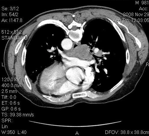
Arterial phase CT shows the right lobe subcapsularhypervascular HCC node in cirrhotic liver Transradial TACE procedure – 5Fr BLK catheter is positioned in CA Selective angiography – microcatheter is positioned in the HCC node feeder branch Selective angiography after DEBDOX (Drug-Eluting Beads loaded with Doxorubicin) depicts complete technical success Follow up arterial phase CT in 3 weeks after TACE shows complete response