-
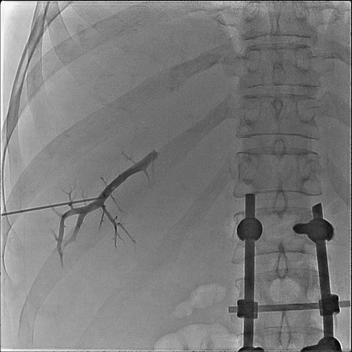
Contrast injection depicts 5th segment PV branch after US guided punctrure
-
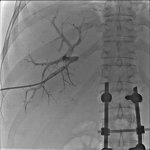
Introducer sheath is positioned and portography performed
-
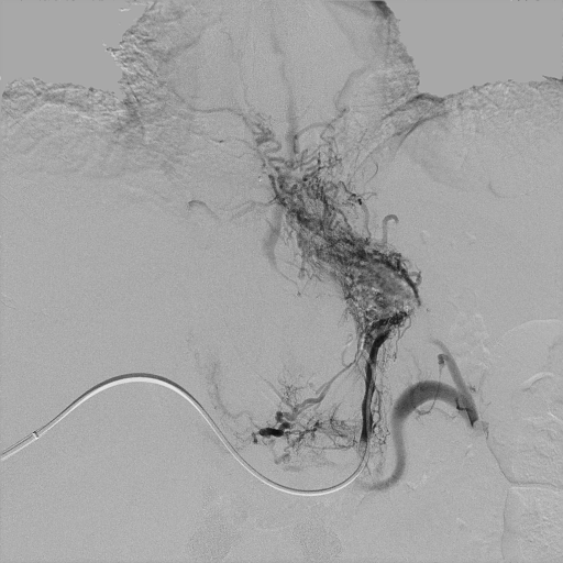
Contrast injection using 5 Fr diameter advantage catheter, positioned in LGV via PV depicts veinsof lower esophagus
-
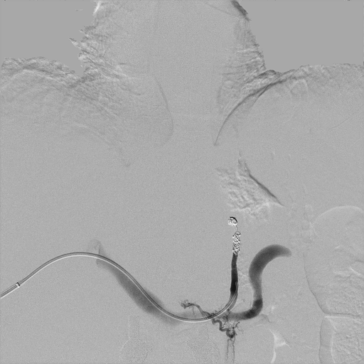
No more esophageal veins are seen after LGV coil embolization